Hydrocephalus (Fluid Accumulation in the Brain)
Hydrocephalus (Fluid Accumulation in the Brain)
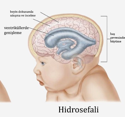
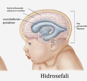
Hydrocephalus is a term derived from the words hydro = water and cephaly = head. It is generally known as excessive fluid accumulation in the brain. The water referred to here is "cerebrospinal fluid." Increased levels of this fluid, found in certain chambers of the brain, can increase intracranial pressure and lead to brain damage.
Cerebrospinal fluid is constantly produced and reabsorbed throughout the day. This fluid surrounds the brain and spinal cord and circulates continuously. It has three primary functions: to reduce the harmful effects of trauma to the brain and spinal cord, to help nourish the brain and transport waste products, and to regulate changes in brain pressure by circulating between the brain and spinal cord.
Hydrocephalus can occur at any age, but it most commonly occurs in children and the elderly (over 60). Approximately one in 500 children develops hydrocephalus. In most of these cases, diagnosis is made at birth, before birth, or in early infancy. Although rare, it can be due to genetic (hereditary) disorders or developmental disorders. Common causes include intracerebral hemorrhages, head trauma, brain tumors, bleeding related to premature birth, and meningitis.
Findings
Symptoms of hydrocephalus vary from person to person. Common symptoms are listed below by age group.
Newborn (0-2 months)
- Excessive growth of the head
- Thinning of the scalp
- The veins on the head become more prominent
- Vomiting, restlessness
- Downward turning of the eyes
- Seizures or inability to communicate
In children (2 months and above)
- Abnormal growth of the head
- Headache, nausea, vomiting, fever
- Double vision, restlessness
- Regression in walking or speaking
- Communication disorder
- Loss of sensory-motor functions, seizures
- Difficulty staying awake or waking up
In middle-aged adults
- Headache
- Difficulty waking up or staying awake
- Balance disorder
- urinary incontinence
- Personality disorder
- Dementia
- Visual impairment
In the elderly
- Communication disorder
- Unsteadiness in walking
- Difficulty remembering
- Headache
- urinary incontinence
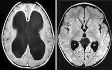
Diagnosis
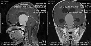
Before starting treatment for a patient with hydrocephalus, your doctor will discuss the condition with you, ask questions, perform an examination, and request certain tests (e.g., brain CT scan, magnetic resonance imaging, and brain ultrasound). The diagnosis of hydrocephalus, its cause, and the treatment plan required will be determined after these tests. A large head alone does not necessarily indicate hydrocephalus in children. However, the diagnosis can be confirmed using brain imaging techniques.
If the diagnosis is made in the womb before the baby is born, a report from the hospital's ethics committee is required for pregnancy termination according to current laws.
Causes of Hydrocephalus
The causes of hydrocephalus vary depending on age group.
1- Newborn (0-2 months)
Congenital:
These patients constitute the largest group. It may occur solely with hydrocephalus or in conjunction with other congenital anomalies of the spine (meningomyelocele).
Intracerebral hemorrhages:
Brain chambers usually expand after spontaneous bleeding.
2- Children and adults
- Brain infections
- Brain hemorrhages
- Brain tumors
- Head traumas
3- Elderly people
Normal pressure hydrocephalus:
It is the enlargement of the brain chambers following decreased absorption of cerebrospinal fluid.
Hydrocephalus Treatment
Hydrocephalus cannot be treated with medication. It can only be corrected through surgical interventions performed by neurosurgeons. The surgical procedures chosen will vary depending on the underlying cause of the hydrocephalus.
If there is an obstruction causing cerebrospinal fluid circulation, surgical treatment can be directed at the cause (tumor, cyst, etc.). If the obstruction cannot be relieved, the intracerebral circulation pathways of cerebrospinal fluid can be surgically altered.
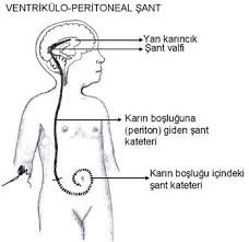
Because restoring cerebrospinal fluid circulation is not possible in most patients, the fluid must be diverted from the brain to another body cavity. A long, thin, elastic, silicone tube called a "shunt" is used for this transfer. A "pump" is located under the scalp to operate in a unidirectional, controlled manner. Excess cerebrospinal fluid is transported to another part of the body through this thin tube, preventing increased pressure within the brain. However, because the brain constantly produces fluid, this system must operate continuously. Because the shunt is located under the skin, it is only noticeable in infants. In children and adults, the tube can be felt under the skin by palpation.
The shunt is surgically implanted under general anesthesia. A small incision is made in the skull, and the tip of the shunt is placed in the chamber within the brain that contains cerebrospinal fluid. A tunnel is then created under the skin of the head, neck, and abdomen, and the other end of the shunt is directed into the heart or abdominal cavity, where the fluid can be easily absorbed. A short course of antibiotics may be used to prevent postoperative infection.
After surgery, the patient is observed in the hospital for a period of time. Symptoms usually resolve after a while. However, if permanent brain tissue damage has occurred, some functions may not recover. The most important reason for failure to recover functions such as vision and intelligence is delayed treatment. The length of hospital stay varies depending on the patient's recovery. These patients require long-term monitoring to ensure the shunt is functioning properly. A significant portion of patients treated for hydrocephalus can lead normal lives. Shunt replacement may be necessary in cases of malfunction or infection.
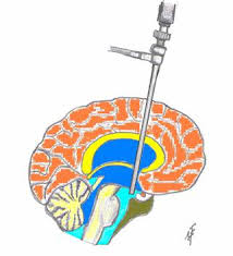
In the surgery called third ventriculostomy, a hole is opened in the skull and with the help of a camera-equipped tip called an endoscope, the adhesions in the brain cavities are opened and the circulation of accumulated water is accelerated.