Spinal Cord Tumors

SPINAL CORD TUMORS
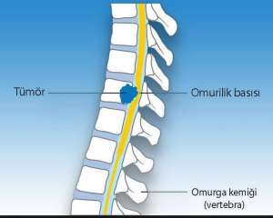
Spinal tumors are tumors located in the spinal cord and nerve roots within the spinal canal. These tumor cells continue to grow uncontrollably and cause harm to the patient.
Spinal tumors can be benign (non-cancerous) or malignant (malignant). Primary tumors refer to tumors originating in the spine or spinal cord itself, while metastatic tumors refer to tumors that have spread from elsewhere in the body to the spine. Spinal tumors can occur in three primary areas: the cervical (neck), thoracic (back), lumbar (lower back), and sacral (tailbone). They are also classified by their location within the spine, such as anterior (front) or posterior (back).
Spinal cord tumors, with their significant morbidity and mortality rates (illness and death), have become a focus of neurosurgery due to the positive outcomes achieved with early diagnosis and appropriate treatment methods. In parallel with technological advances, increased diagnostic capabilities and the development of surgical techniques, particularly microsurgery, have led to increased treatment success rates.
Approximately to of central nervous system tumors are located in the spinal cord. The prevalence of spinal tumors in the general population varies between 2 and 10 per 100,000. Differentiating spinal tumors by location facilitates diagnosis and treatment. Therefore, the relationship with the dura mater (the outer layer of the spinal cord membrane) is taken into account in classification. Spinal tumors are classified accordingly as extradural, intradural extramedullary, and extramedullary. The ratio of intradural to extradural is 2:3. Of all spinal tumors, are extradural, are intradural extramedullary, and %5 are intradural intramedullary.
Metastatic tumors constitute the majority of extradural tumors. In addition to metastatic tumors, primary spinal tumors are also included in extradural tumors. Neurofibroma and meningioma constitute the majority of intradural extramedullary spinal tumors. Ependymoma, astrocytoma, and hemangioblastoma constitute the majority of intradural intramedullary spinal tumors.
Spinal tumors are predominantly benign. Improvements in early diagnosis and treatment have led to improved outcomes.
Treatment is mainly aimed at total removal of the tumor by surgery, and most tumors are generally suitable for such a treatment method.
General Clinical Findings and Diagnostic Methods
Both benign and malignant tumors present with pain in the back or lower back that is not related to movement. This pain is usually not caused by trauma, exercise, or stress. However, the pain increases with exercise and worsens at night. The pain can radiate to the hips, legs, and arms if the tumor is in the neck, and conservative treatment is not effective.
While the patient's symptoms and signs vary depending on the tumor's location, they generally occur when the tumor grows and presses on the spinal cord, nerve roots, or blood vessels. Compression of the spinal cord by itself is life-threatening. Additional symptoms are listed below:
- Weakness or numbness in arms and legs
- Difficulty in walking, which causes the patient to fall
- Loss of pain and temperature sensation
- Impaired bowel and bladder control
- Paralysis can occur in varying degrees.
- Scoliosis or other spinal deformities may be the result of large but benign tumors.
DIAGNOSIS
The first step in diagnosis begins with the patient's history and examination. Radiological diagnosis is performed to confirm this. A thorough neurological examination can help pinpoint the tumor's location.
Direct X-ray
This X-ray reveals the bone structure. It can only aid in diagnosis if there is a bone lesion, but it cannot determine whether there is an infection or a tumor.
Computed Tomography (CT)
With this imaging, diagnosis can be made, the canal diameter can be measured and it shows the bone structure very well.
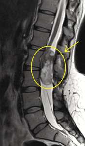
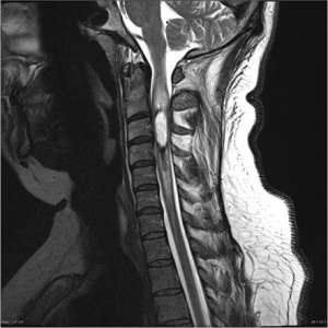
Magnetic Resonance Imaging (MRI)
Using computer technology and a magnetic field, a 3D image of the body can be created. Unlike other methods, MRI provides a very clear view of the nerve roots, spinal cord, surrounding soft tissue, and tumors.
Once the tumor is identified, definitive determination of whether it is benign or malignant is made by taking a sample of the tumor and examining it under a microscope. If the tumor is malignant, pathology can also reveal its type.
If it is metastasis, primary investigation should be performed, abdominal USG, Tomography and similar tests can be performed.
Treatment Options
Surgical Treatment
Treatment primarily aims to remove the entire tumor (total) through surgery, and most tumors are generally amenable to this treatment. The indication for surgery depends on the tumor type. Primary spinal tumors should be completely removed if possible to ensure a possible cure. The goal for metastatic tumors is to relieve pain, increase spinal stability, and treat or prevent neurological deterioration. Surgical treatment for metastases is recommended if the patient's life expectancy is greater than 12 weeks and the tumor is resistant to radiotherapy and chemotherapy. Other indications include pain resistant to medication, poor spinal stability, and compression of the spinal cord.
Surgical Approaches:
- Posterior approach:With a posterior approach, the nerve roots and dura mater membrane are seen.
- Anterior approach:Tumors located anteriorly are more easily controlled with the frontal approach.
- 360-degree approach:Both anterior and posterior approaches can be performed in the same session.
CyberKnife Radiosurgery
Radiosurgery has seen significant advances in recent years. CyberKnife radiosurgery has been beneficial in pain control and improving quality of life. Short treatment times, rapid recovery, and a positive response to treatment are among the primary benefits of CyberKnife radiosurgery. This technique can be used primarily for spinal lesions, but can also be used in inoperable cases, in patients who have previously received radiotherapy, or as a supplement to surgical techniques.